Fig. 3.

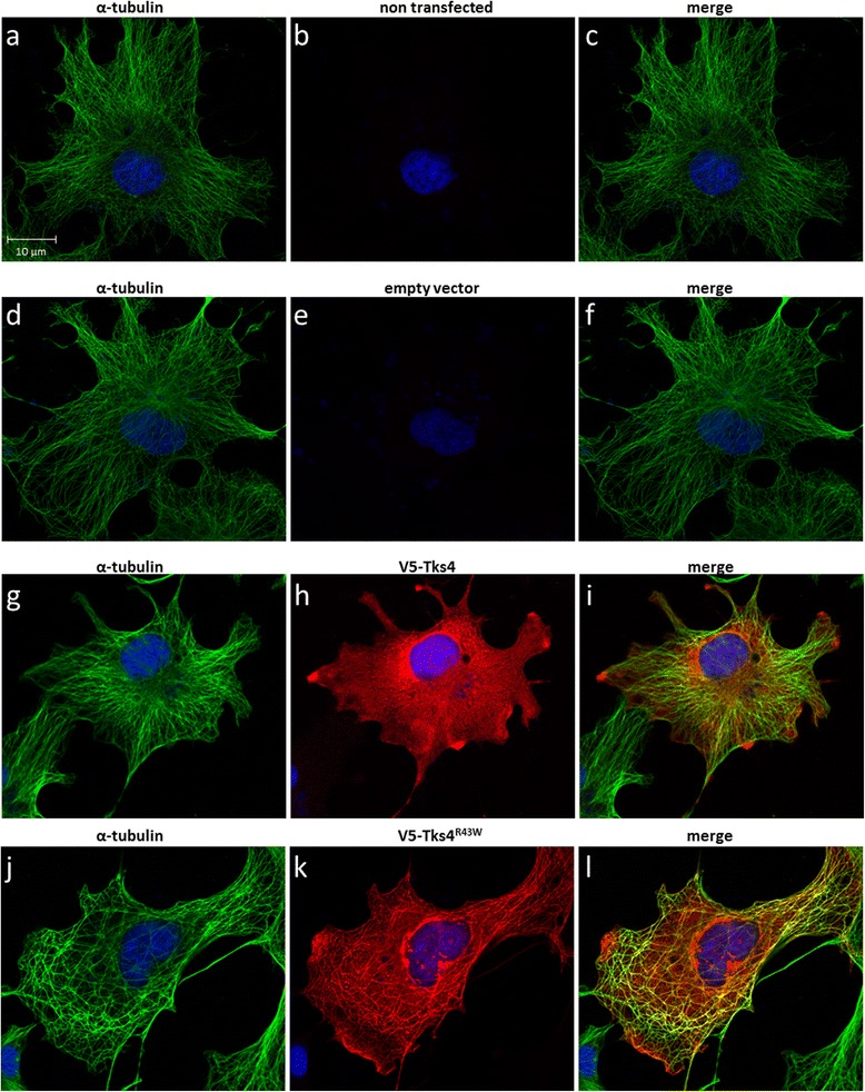
The Tks4R43W mutant protein colocalizes with the microtubule network of the cytoskeleton. COS7 cells were transiently transfected with wild type V5-Tks4 (3g, h, i), V5-Tks4R43W (3j, k, l) or V5-empty (3d, e, f) constructs. Non-transfected cells are also shown (3a, b,c). After 18 h, cells were fixed and stained for Tks4 (with anti-V5 antibody, red, 3b, e, j, k) and α-tubulin (green, 3a, d, g, j). Merged pictures are also shown (3c, f, i, l). Cell nuclei were visualized by DAPI staining. The scale bar represents 10 μm
