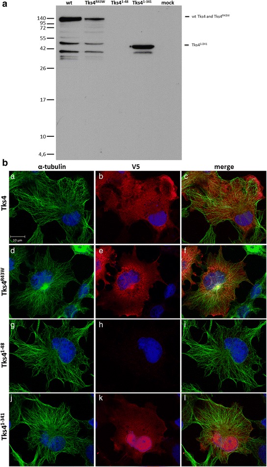
Fig. 8.

The expression patterns of Tks4 mutant proteins. 8a, COS7 cells were transiently transfected with V5-Tks4, V5-Tks4R43W, V5-Tks41–48 and V5-Tks41–341 constructs. After 18 h the cells were lysed, the lysates were separated by centrifugation and the supernatant analyzed by Western blotting for Tks4 with anti-V5 antibody. 8b, COS7 cells were transiently transfected with V5-Tks4, V5-Tks4R43W, V5-Tks41–48 and V5-Tks41–341 constructs. After 18 h, cells were fixed and stained for Tks4 (with anti-V5 antibody, red) and α-tubulin (green). Cell nuclei were visualized by DAPI staining. The scale bar represents 10 μm
