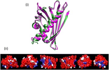
Figure 2.

Structural homology of Fag s 1. (i) Superimposed Ribbon drawing of the Fag s 1 (green) homology model superimposed onto the template crystal structure of Bet v 1 major allergen (magenta) (ii) Space filling model of Fag s 1; conserved amino acid residues are coloured in red, different residues in white, homologous substitutions in blue (69% identity/80% similarity).
