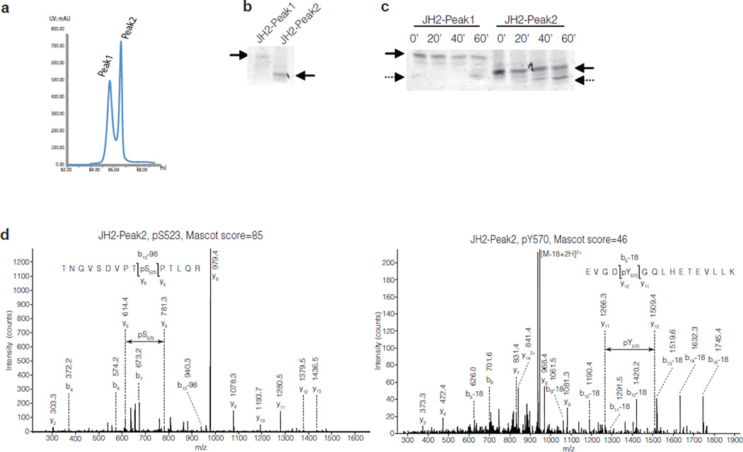
Figure 2.
Identification of phosphorylated residues in JAK2 JH2. (a) Chromatogram of JAK2 JH2 purification showing the peaks from anion-exchange chromatography. (b) Coomassie staining of a native-gel electrophoresis of JH2-Peak1 and JH2-Peak2 proteins. (c) Coomassie staining of a native-gel electrophoresis of purified JH2-Peak1 and JH2- Peak2 after kinase reaction. (d) MS-MS spectra of the phosphorylated residues in JAK2 JH2-Peak2 4h kinase assay. Left: JH2-Peak2 is stoichiometrically phosphorylated at Ser523. Right: JH2-Peak2 is partially phosphorylated at Tyr570.

