
Figure 3.
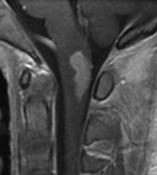
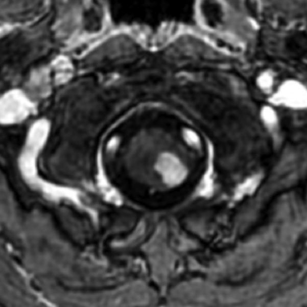
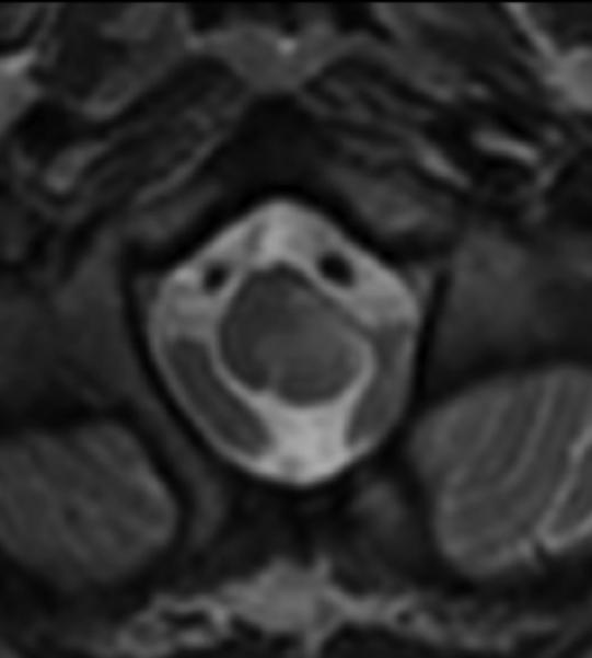
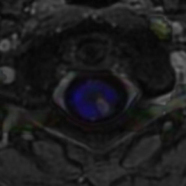
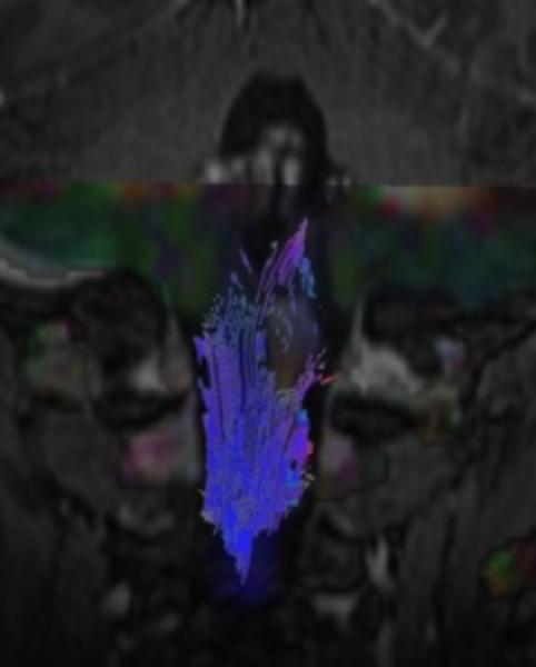
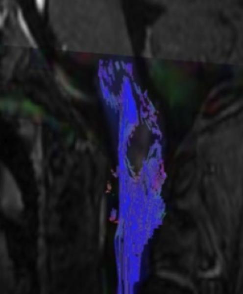
Sagittal (A) and axial (B) T1W image of the upper cervical cord shows an enhancing intramedullary mass in the left dorsolateral aspect of the spinal cord immediately caudal to the foramen magnum. Axial T2W image (C) at the superior aspect of the lesion shows expansion of the left dorsolateral aspect of the upper cervical cord with T2 prolongation extending beyond the margins of the enhancement. Axial T1W post-gadolinium image at the same level with DE-FA overlay (D) shows diminished FA in the left dorsolateral aspect of the cord, corresponding to the expected location of the spinal trigeminal tract and causing the ipsilateral jaw pain. Coronal (E) and sagittal (F) T1W post-contrast images with DT-FT overlay show disruption of fibers by the mass.