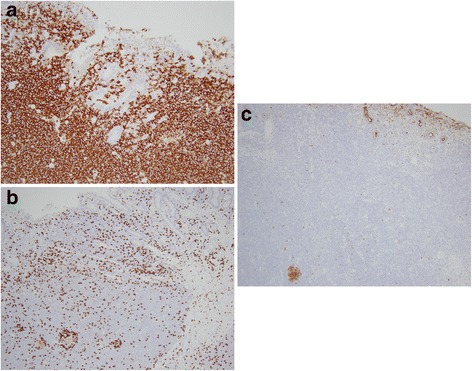
Fig. 3.

a Lymphocytic infiltrate consists predominantly of CD20 positive B-cells. CD20 immunohistochemical stain. 200x. b B-cells are negative for CD5 which highlights scattered T-cells. CD5 immunohistochemical stain. 200x. c B-cells are predominantly negative for CD10 which highlights few residual germinal center B-cells. CD10 immunohistochemical stain. 200x
