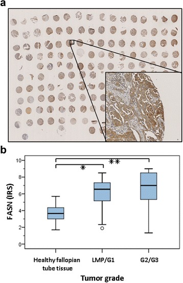
Figure 1.

Immunhistochemical analyses of fasn protein expression in patient material. Immunhistochemical analyses of FASN protein expression in a TMA comprising formalin-fixed, paraffin-embedded samples of ovarian cancers of different grades (6 LMP, 9 G1, 42 G2, and 47 G3 tumors) and histological subtypes (serous papillary, mucinous, or endometrioid) from 104 patients versus in 12 healthy fallopian tissue samples. (a) Representative TMA slide immunohistochemically-stained with FASN antibody showed strong FASN expression in ovarian cancer. (b) Statistical evaluation of FASN expression applying the immunoreactive score (IRS), which incorporates protein staining intensity and the percentage of protein-positive cells. Statistically significant FASN overexpression was proven for LMP/G1 tumors or G2/G3 tumors vs. normal tissues (respectively *P < 0.005 and **P < 0.001, U test).
