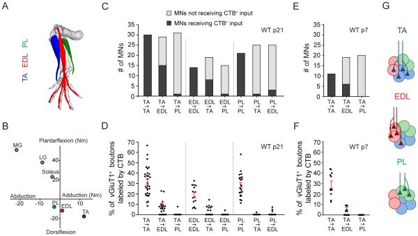
Figure 1. Patterns of Heteronymous Sensory-Motor Connections in the Anterolateral Crural Synergy Group.
(A) Schematic of anterolateral crural muscle anatomy. TA = tibialis anterior. EDL = extensor digitorum longus. PL = peroneus longus. Image generated using The Mouse Limb Anatomy Atlas (Delaurier et al., 2008).
(B) Lines of action of muscles in cat. Axes represent torques (in newton-meters) evoked by individual muscle nerve stimulation. Posterior crural muscles shown in grey. MG = medial gastrocnemius. LG = lateral gastrocnemius. Adapted from (Nichols, 1994).
(C) Number of self and synergist motor neurons innervated by sensory afferents from a given muscle in p21 wild-type mice (n = 14-30 MNs; 3-7 mice per pair).
(D) Density of sensory synaptic connections with self and synergist motor neurons in p21 wild-type mice. Each point represents one motor neuron (n as in (C)). Red lines indicate mean ± SEM for motor neurons receiving input.
(E) Number of self and synergist motor neurons innervated by TA sensory afferents in p7 wild-type mice (n = 11-20 MNs; 2-3 mice per pair).
(F) Density of TA sensory synaptic connections with self and synergist motor neurons in p7 wild-type mice (n as in (E)).
(G) Schematic depicting organization of sensory-motor connectivity within the anterolateral crural synergy group.
All data reported as mean ± SEM. For related data, see also Figure S1.

