Figure 1.

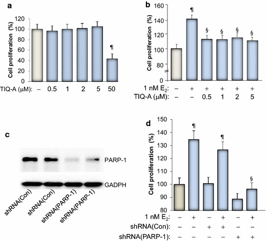
Effect of PARP inhibition on E2-stimulated growth of MCF-7 cells. a MCF-7 cells were treated for 48 h in the absence or presence of increasing concentrations of TIQ-A. Cell viability was assessed using a MTT assay. ¶Difference from viability values of cells that did not receive TIQ-A; p < 0.05. b MCF-7 cells were stimulated with 1 nM E2 for 48 h in the absence or presence of the indicated TIQ-A concentrations. Cell viability was then assessed using a MTT assay. ¶Difference from viability values of cells that did not receive TIQ-A; p < 0.05. §Difference from viability values of cells that were treated with E2 alone; p < 0.05. c MCF-7 cells were transduced with a viral vector encoding control shRNA or a shRNA targeting human PARP-1. Protein extracts were prepared and subjected to immunoblot analysis with antibodies against PARP-1 or GAPDH. d Cells expressing control or PARP-1-targeting shRNA were treated with E2 for 48 h after which viability was assessed as described above. ¶Difference from respective untreated controls; § difference from E2-treated cells; p < 0.05.
