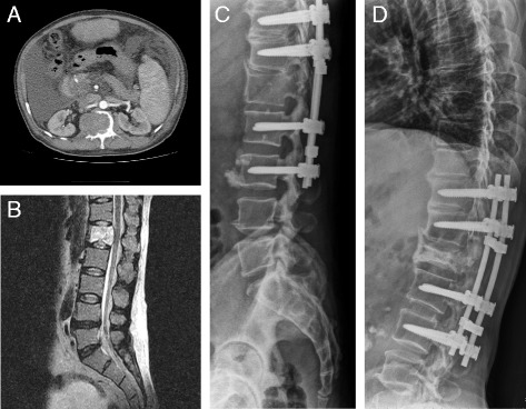
Fig. 2.

Preoperative and postoperative images of a 49-year-old man with L1 vertebral metastatic spinal cord compression from pancreatic cancer who underwent surgery using the posterolateral transpedicular approach without anterior vertebral reconstruction. The CT angiography (a) and MRI image (b) demonstrate metastatic spinal tumor with cord compression at L1. The immediate postoperative radiographs (c) and the 28-month follow-up radiographs (d) demonstrate no screw loosening or implant failure
