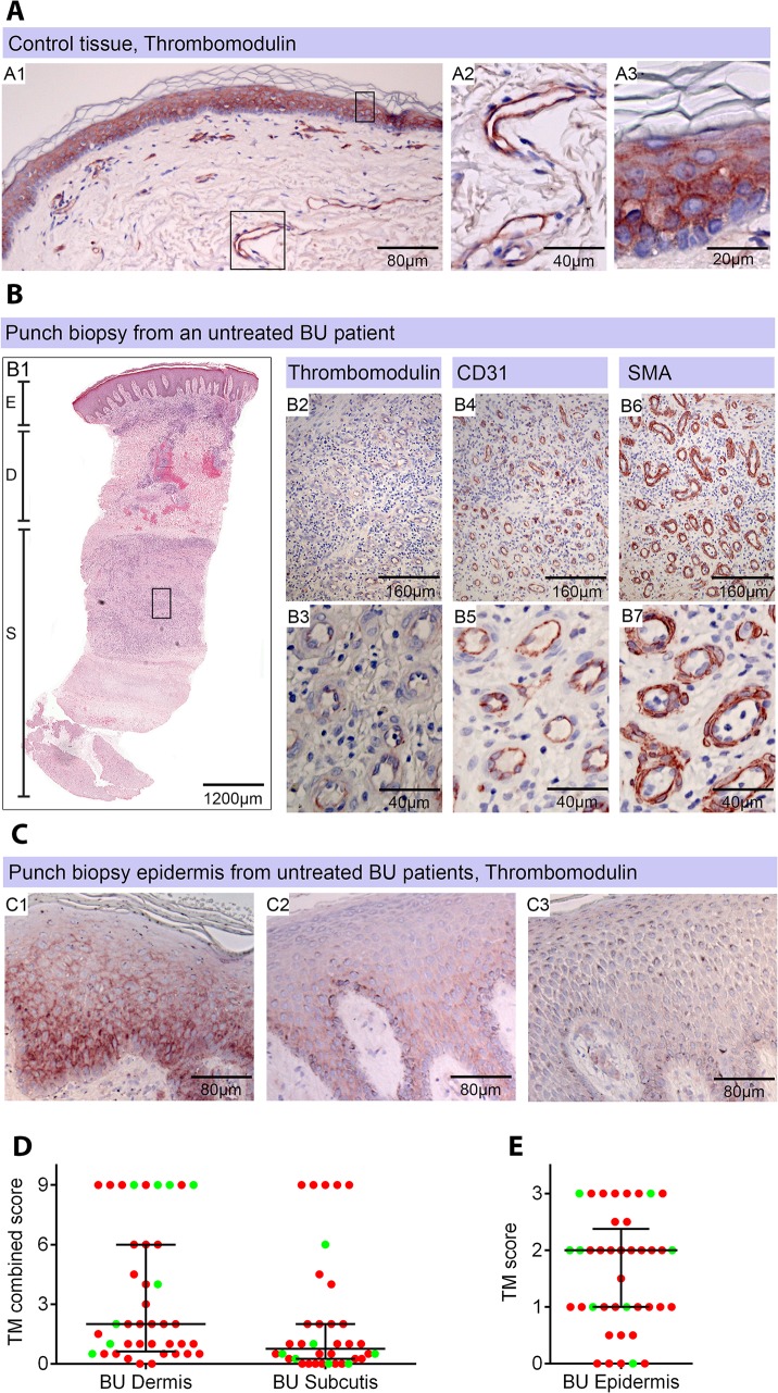
Fig 4. Thrombomodulin (TM) expression is dysregulated in Buruli ulcer patient skin and tissue.
Histological sections were stained with α-TM, α-CD31 (PECAM-1) or α-SMA (smooth muscle actin) antibodies and counterstained with Haematoxylin or with Haematoxylin-Eosin (B1). Slides were analyzed with a DM2500 Microscope (Leica). Pictures were taken either with an Aperio scanner (B1) or a Leica DFC 420 camera and the Leica application Suite V4 software. Comparative staining on healthy skin sample from an unaffected donor (A) or 4mm punch biopsies from laboratory confirmed BU patients (B and C). Typical results are shown. A. TM staining of healthy skin. A1, Low magnification image; A2 and A3, higher magnification showing strong TM staining of endothelial cells and keratinocytes, respectively. B1. Scan of a HE stained BU punch biopsy (E; Epidermis, D; Dermis, S; Subcutis). B2 –B7, higher magnification of subcutaneous tissue showing reduced TM staining in the endothelium (B2 and B3). This patient had a combined score of 2 in the dermis (coverage 2, intensity 1) and 2 in the subcutis (coverage 2, intensity 1). Endothelial cells in the region still showed reasonable staining for CD31 (B4 and B5) and strong staining for αSMA (B6 and B7), scoring 2.5 in the dermis and 3 in the subcutis. C. Higher magnification of the epidermis showing variable reduction in TM staining in the keratinocytes of BU patient skin, ranging from reduced (C1) to no staining (C3). Hyperplasia of the epidermis, as seen in these three patients, is typical of BU. D and E. TM staining in the dermis, subcutis and epidermis was scored as described in Materials and Methods. The score for each individual biopsy analysed is shown, consisting of 31 patients with ulcers (red circles) and 9 patients with plaque lesions (green circles). In all cases, error bars show the median and 25–75% percentile scores for the all BU patients. D. Scores of dermis and subcutis are for endothelial TM and were obtained by multiplying two scores (each 0–3) of intensity of staining and the coverage, (healthy tissue scored the maximum 9). Numbers for subcutical staining are slightly lower since those patients that had no intact endothelium in the subcutis according to SMA staining were excluded. E. Score for epidermal TM (keratinocytes) are relative to a maximum of 3 (intensity only).

