
Fig 3. Ligand displacement assay of HSA with Quercetin, 4PBA.
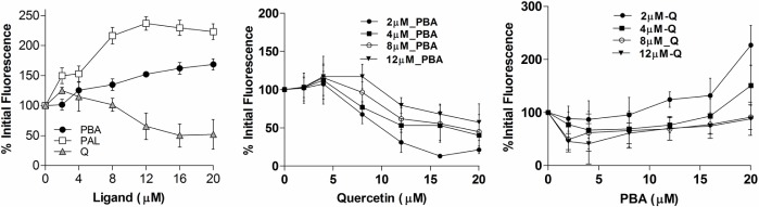
a) Percentage initial fluorescence of HSA (at 345 nm) upon addition of 4PBA, Palmitic acid and Quercetin (2–20 μM). b) Binding of 4PBA and Quercetin at different sites. Tryptophan fluorescence of HSA was monitored at 345 nm in the presence of 4PBA. To HSA-PBA complex (of varying PBA concentration: 2–12 μM), Quercetin was added from 2–20 μM and c) To HSA-Quercetin complex (of varying Quercetin concentration: 2–12 μM), PBA was added from 2–12 μM. HSA fluorescence was normalized to 100% in the absence of added ligands.