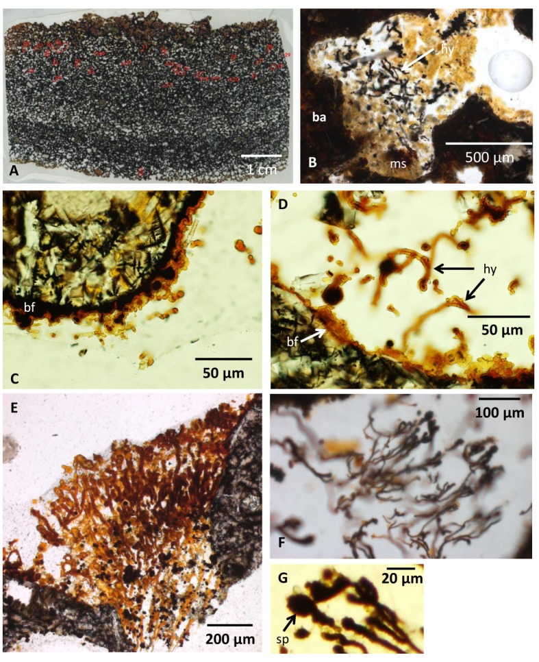
Fig 2. Microphotographs of the vesicular basanite and fungal communities within.

(A) Thin section showing the vesicularity of the pillow lava. (B) A vesicle partly filled with marine sediments. Fungal hyphae occur in association with the sediment. (C) Fossilized biofilm lining the vesicle walls with spherical structures and a few protruding filaments. (D) Fossilized biofilm on the vesicle wall from which hyphae protrude. (E) Hyphae of the first bush-like type with a directed growth occupying the void in between two vesicles. (F) Hyphae of the first bush-like type with a directed growth and oval terminal swellings. (G) Close-up of oval terminal swellings. Legend for all figures: ba, basanite; ms, marine sediments; hy, hyphae; bf, biofilm (fossilized); sp, sporophore/spore; an, anastomose; cs, central strand; zph, zygophore; pr, progametangia; ga, gametangia; mzy, maturing zygote; zy, zygote; di, diatome, ra, raphe; ar, areolaes; fmo, fragmented marine organism; ha, haustorium.
