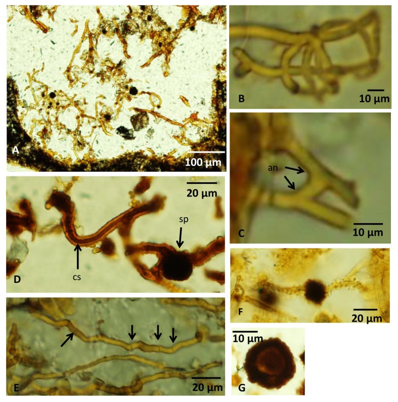
Fig 3. Microphotographs of fungal hyphae and spores.

(A) Overview of a vesicle filled with hyphae of the second type without a mutual directed growth. (B) Branched hyphae. (C) Hyphae with an anastomose. (D) A hyphae with a central strand. A spherical spore structure is seen on another hypha. (E) Irregularly spaced septa along a hyphae marked with arrows. (F) A dark, swollen, anisotropic spore structure along a hyphae. (G) A spherical spore with a distinct core with carbonaceous matter according to Raman analyses. For legends see caption of Fig 2.
