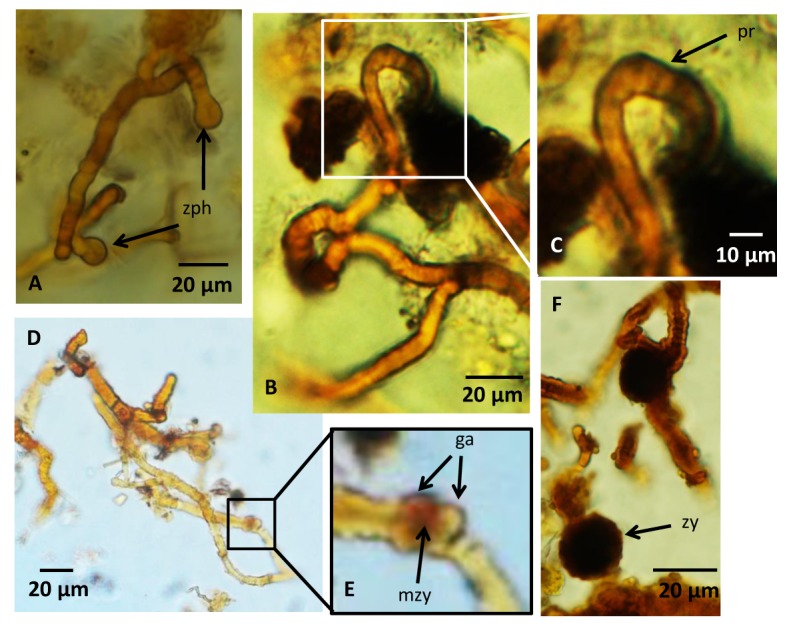
Fig 4. Microphotographs of zygospores and a Zygomycetes reproduction cycle.

(A) Two zygophores approaching each other. (B,C) Fusion of two zygophores to form progametangia. Note how the zygophores are tightly appressed parallel to each other while fusing to form the progametangia. (D,E) Gametangia are separated by septa and a structure develops in between them that could represent early stage of zygote formation with fusion wall beginning to degrade. (F) Image showing a swollen, dark structure with a warty appearance that represents the last stages of the zygospore formation, the zygote. For legends see caption of Fig 2.
