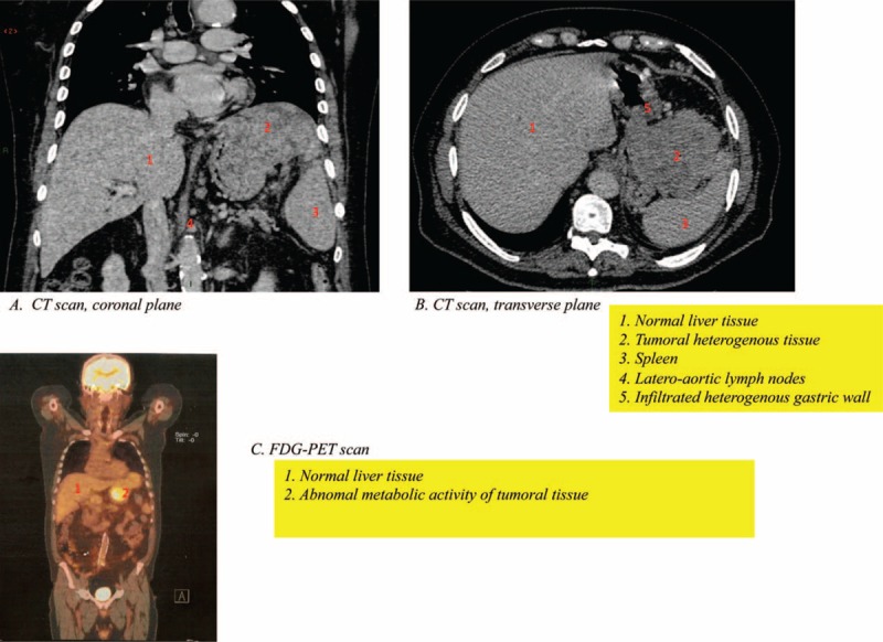
FIGURE 1.

Imaging at diagnosis showing at CT scan (A and B) a large heterogeneous tumor measuring 10 × 8 mm initially the origin was taken for a gastric lesion. The tumor extends to the diaphragm, the splenic hilum, and lymph nodes. The liver seems to be normal with no evident lesions. The positron emission tomography scan showed abnormal metabolic activity located near the liver and the spleen hilum (C). CT = computed tomography.
