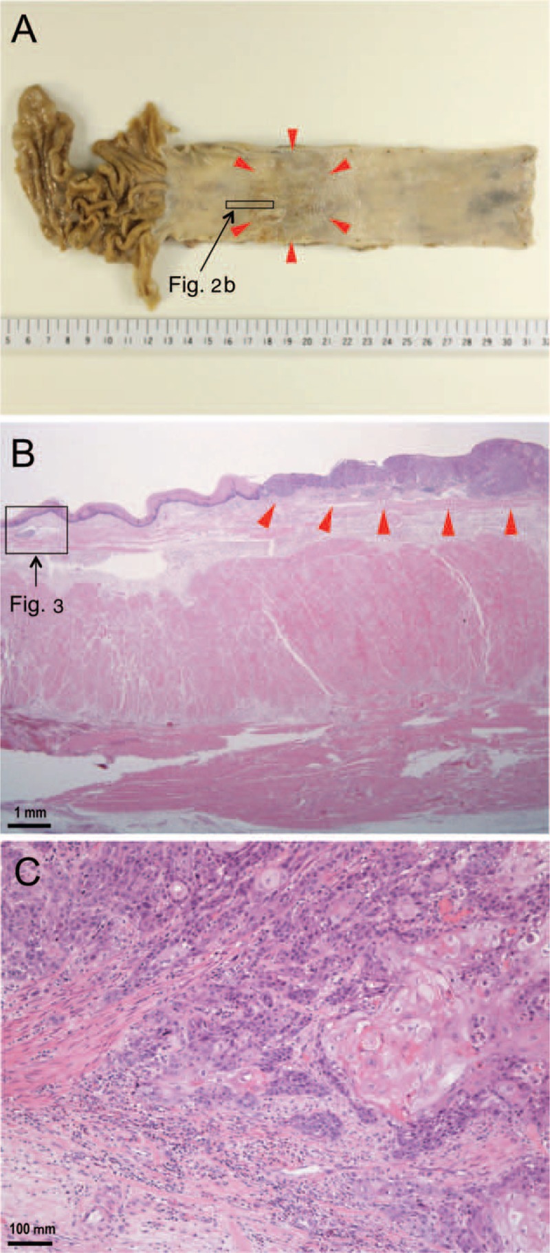
FIGURE 2.

Pathological findings for the examined esophageal cancer. A: A gross image depicts a superficial (0 – Ip + IIc type) single tumor 47 × 38 mm in size located in the middle to the lower thoracic portion of the esophagus (arrowheads). B: A low-power photomicrograph reveals a primary lesion (arrowheads) and an area of retrograde lymphatic invasion distant from the primary lesion (rectangle) (hematoxylin and eosin stain). C: A high-power photomicrograph illustrates the primary lesion and features indicating the invasion of well-differentiated squamous cell carcinoma into the submucosal layer (hematoxylin and eosin stain).
