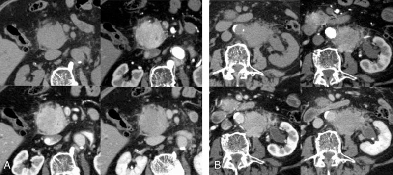
FIGURE 1.

(A) Multiphasic contrast-enhanced abdominal CT demonstrating a solid mass of 5 cm in diameter with homogenous intravenous contrast enhancement in the arterial phase in the head of the pancreas. Upper left column: plain CT; upper right column: arterial phase; lower left column: portal venous phase; and lower right column: delayed phase. (B) Multiphasic abdominal CT of demonstrating a 4.5-cm solid mass in the retroperitoneum. Upper left column: plain CT; upper right column: arterial phase; lower left column: portal venous phase; and lower right column: delayed phase. CT = computed tomography.
