
FIGURE 2.
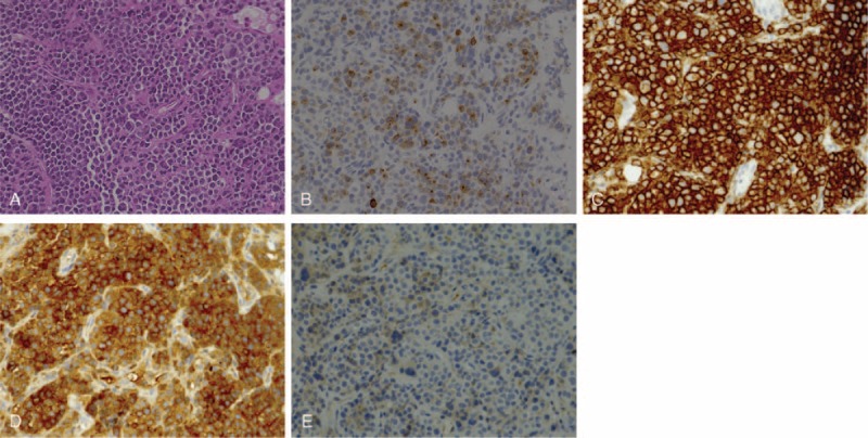
The pathological findings of a CT-guided biopsy of the retroperitoneal mass. (A) Hematoxylin-eosin staining. Multiple sheets of atypical plasma cells with irregular nuclei were located eccentrically in the cytoplasm. (B) CD79a. (C) CD138. (D) Immunoglobulin κ chain. (E) Immunoglobulin λ chain. The immunohistochemical profile of the tumor was positive for CD79a, CD138, and the κ light chain, which was consistent with plasmacytoma. CT = computed tomography.