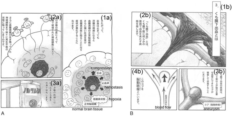
FIGURE 2.

These pages explain the pathogenesis. (A) Explains how intracerebral hematoma destroys perihematomal neurons and (B) the image of how aneurysms rupture. Translation: (1a) Therefore, although it stops bleeding, symptoms will worsen during periods of perihematomal edema, which compress normal tissue strongly and make it hypoxic. (2a) Perihematomal edema kills more normal perihematomal tissue. (3a) We administer patients an intravenous drip injection, which improves brain edema during periods of perihematomal edema. (1b) What is subarachnoid hemorrhage? (2b) A subarachnoid hemorrhage is defined as bleeding into the subarachnoid space for some reason. (3b) This may occur usually from a ruptured blood-filled balloon-like bulge in the wall of a blood vessel. (4b) We call it an aneurysm.
