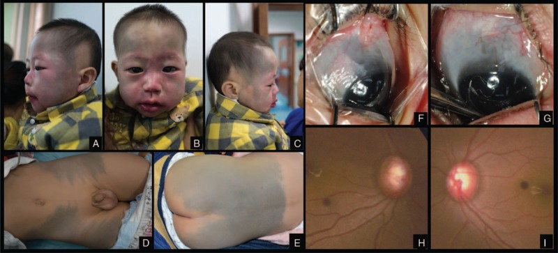
FIGURE 1.

Case 1. Facial port-wine stains distributed along the branches of trigeminal nerve. Gray-blue patches (Mongolian spot) spread over forehead, temporal. Triangular alopecia existed symmetrically on bilateral temporal scalp (A, B, C). Gray-blue patches (Mongolian spot) spread over thigh (D). Gray-blue patches (Mongolian spot) spread over waist and breech (E). Scleral malanocystosis on the right eye (F). Scleral malanocystosis on the left eye (G). Enlarged optic disc cup on the right eye (H). Enlarged optic disc cup on the left eye (I).
