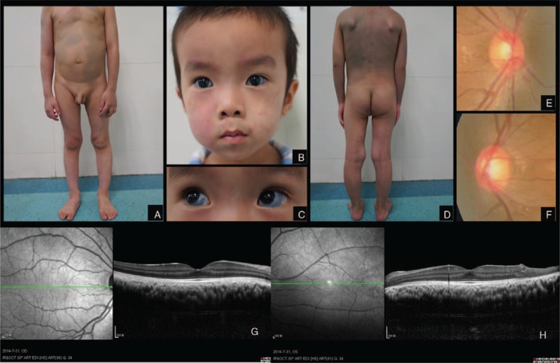
FIGURE 2.

Case 2. Port-wine stains distributed along chest, arms, and legs. Greyish blue patches (Mongolian spots) spreading over abdomen (A). Port-wine stains were found on the face along the 3 branches of trigeminal nerve (B). Bilateral scleral malanocystosis (C). Port-wine stains distributed along back, arms, and legs. Mongolian spots spreading over back (D). Enlarged optic disc cup on the right eye (E). Enlarged optic disc cup on the left eye (F). Increased choroidal thickness in the right eye on OCT scan (G). Increased choroidal thickness in the left eye on OCT scan (H).
