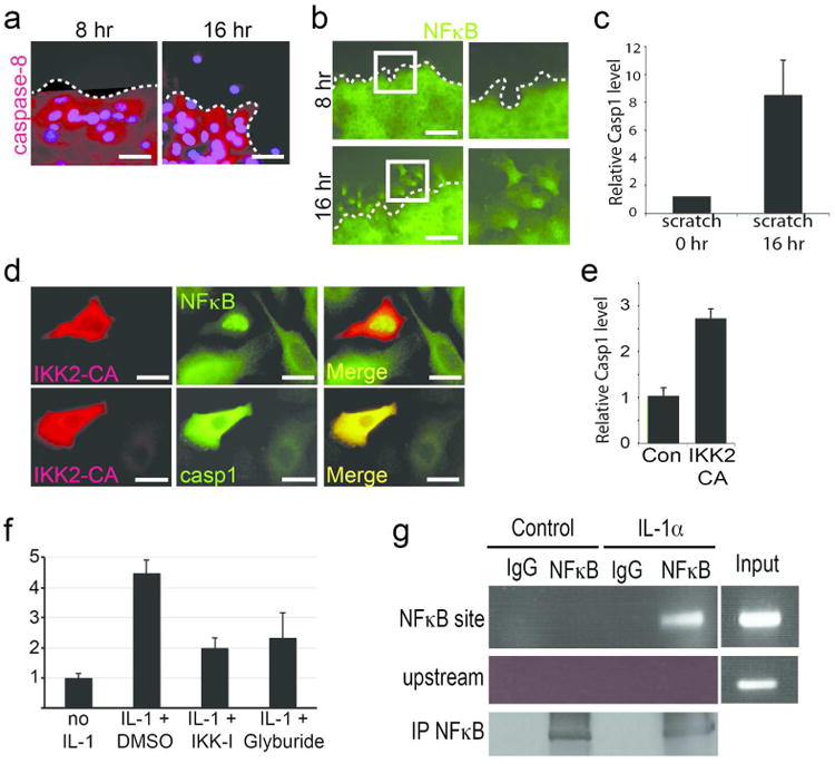
Figure 2. Regulation of caspase-1 by NFκB in wounded cells.

(A) Immunofluorescence of (A) caspase-8 (red), DAPI (blue) and (B) NFκB (green) in primary keratinocytes after 8 or 16 hours of applying a scratch wound (dotted line denotes wound margin). Right panels in (B) are the magnified views of the boxed areas in the left panels. (C) qPCR of caspase-1 RNA levels at 0 and 16 hours after applying scratch wound. (D) Primary keratinocytes were transfected with constitutively active HA-tagged IKK2 (IKK2-CA) and stained for active IKK2 (red) and NFκB (green) or caspase-1 (green). Cells expressing IKK2 have nuclear NFκB and higher levels of caspase-1. (E) qPCR of relative levels of caspase-1 RNA in keratinocytes transfected with empty vector (Con; normalized to 1) or IKK2-CA. (F) Relative caspase-1 protein levels in keratinocytes exposed to IL-1 and DMSO, IKK-I, or glyburide. Caspase-1 protein levels in cells without IL-1α stimulation is normalized to 1 and data are presented as fold difference. Data is the mean of 3 biological replicates and error bars are SEM. (G) ChIP of NFκB upon stimulation with IL-1α and immunoprecipitation with IgG control antibody or an antibody recognizing the p65 subunit of NFκB. * = p<.001. For qPCR experiments (C & E), results are from 3 biological replicates with each PCR reaction performed in triplicate (error bars are SEM).
