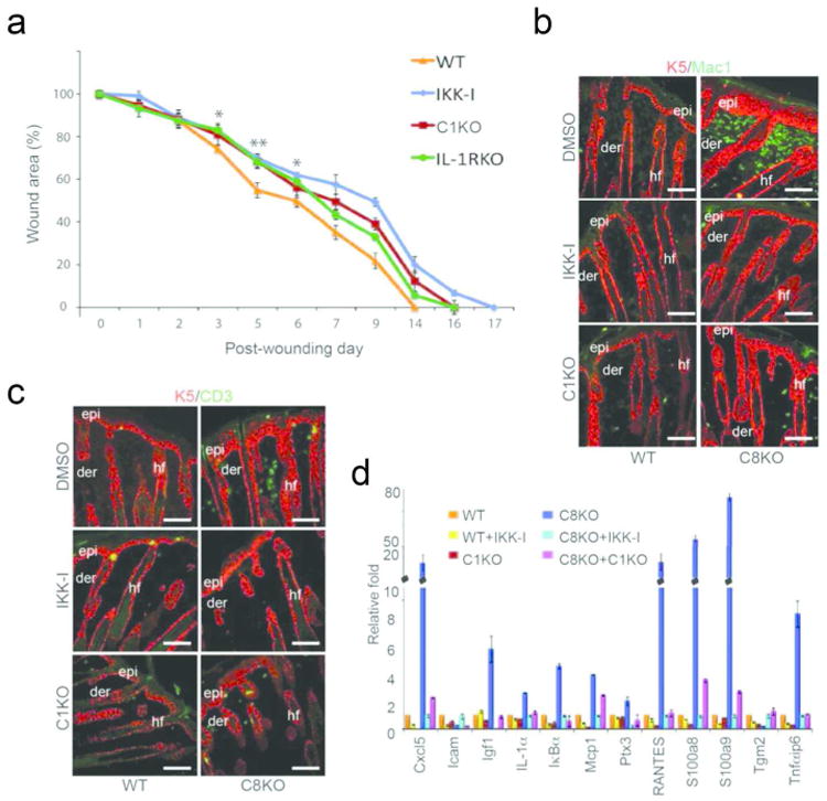
Figure 5. Effect of NFκB and caspase-1 on wound closure.

(A) Rates of wound closure were measured in wild type mice treated daily with DMSO or IKK-I, or in mutant mice lacking either caspase-1 (C1KO) or IL-1 receptor (IL-1RKO). Data points are averages of 6 mice with two wounds/mouse (* denotes p<.001, and ** denotes p<.00001 when comparing DMSO vs. IKK-I kinetics in wild type mice). (B) Recruitment of macrophages (green) in the wild type and caspase-8 null skin treated with DMSO (control) or IKK-I or in skin lacking caspase-1 and -8. (C) Recruitment of T-cells (green) monitored using the pan T cell marker CD3. The epidermis (epi) and hair follicles (hf) in B & C are noted in red with an antibody detecting keratin-5. Scale bar: 50 μm; der = dermis (D) Inflammatory gene expression in the wounded/wound-like skin. RNA was extracted from the wounded skin of wild type mice (treated with DMSO [orange bars] or IKK-I [yellow bars]) or caspase-1 KO mice (C1KO; red bars) 3 days post excisional wounding. RNA was also extracted from the skin of P5 caspase-8 knockout (C8KO) mice (treated with DMSO [blue] or IKK-I [light blue]) or the caspase-8/caspase-1 (C1KO) double knockout mouse [magenta]. Results are from 3 biological replicates for each mouse genotype/treatment and each sample was repeated in triplicate. Error bars are SEM.
