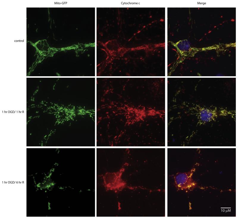
Fig. 5.
Oxygen glucose deprivation results release of cytochrome c from the mitochondria. Mitochondrial targeted GFP transfected primary neurons (column 1) were subjected to 1 h of OGD followed by 1 or 6 h of reoxygenation in glucose rich media. Following fixation, an antibody directed against cytochrome c (red; column 2) was used to identify localization. The yellow signal in the merged overlay indicates co-localization (column 3). In control cells (row 1), cytochrome c immunofluorescence had absolute overlay with mitochondrial GFP signal as seen in the merged column. Following 1-h OGD and 1-h reoxygenation in glucose rich media (row 2), mitochondrial morphology differed from the controls, however, cytochrome c signal co-localized with mitochondria. However, 1-h OGD followed by 6-h reoxygenation (row 3) resulted in widespread cytochrome c signal in the cytoplasm, with fragmented mitochondria. (For interpretation of the references to colour in this figure legend, the reader is referred to the web version of this article.)

