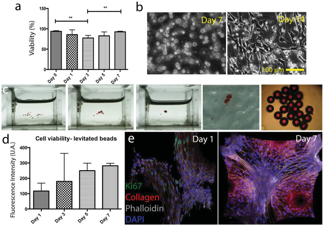
Figure 5.
Evaluation of the effects of levitation and Gd3+ medium on viability and proliferation. a) Long term viability results of 50 × 10−3 m Gd3+ treated hydrogel for 10 min. Lines connecting individual groups indicate statistically significant difference. One-way ANOVA with Tukey's post-hoc tests, *p < 0.05, **p < 0.01. b) Brightfield images of NIH3T3 mouse fibroblasts encapsulating 5 w/v% GelMA hydrogels after 7 and 14 d. c) 3T3 seeded microbeads were self-assembled in 50 × 10−3 m Gd3+ solution with magnetic levitation setup (total exposure time is 10 min). After levitational self-assembly and draining the suspension media out, beads were cross-linked with GelMA for stabilization. d) Cell viability results with Alamar Blue assay. Fluorescent intensity is given as a function of incubation days indicating the increase in biological activity, viability, and cell growth. e) Immunocytochemistry staining of cells seeded on assembled beads. Cell proliferation (ki67 green) and collagen secretion in a week after the levitation.

