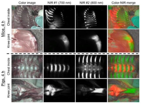
Figure 3.
Dual-channel in vivo fluorescence imaging of cartilage and bone tissues by using C700-OMe and P800SO3− [15] in the same animals. 10 nmol and 1 μmol of C700-OMe and P800SO3− were intravenously injected into 25 g CD-1 mice (top; 0.4 mg kg−1) and 35 kg Yorkshire pigs (bottom; 0.02 mg kg−1), simultaneously, 4 h prior to imaging. All NIR fluorescence images for each condition have identical exposure times and normalizations. Scale bars = 1 cm. Images are representative of n = 3 independent experiments. Pseudo-colored red and green colors were used for 700 nm and 800 nm channel images, respectively, in the color-NIR merged image.

