
Figure 4. Regions comprising the Brain signature 2.
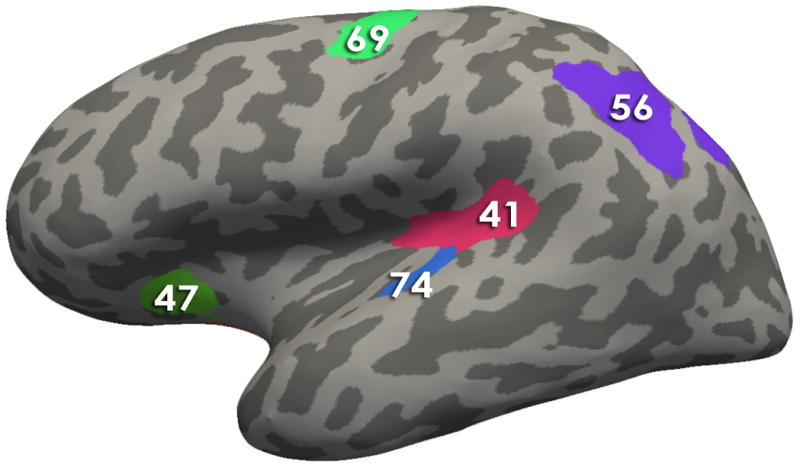
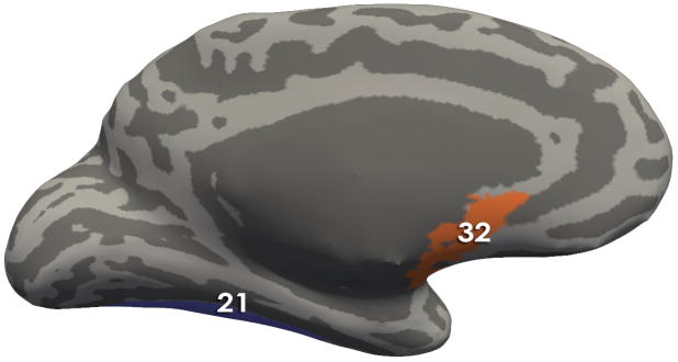
Using Freesurfer, all regions comprising the signatures are depicted on an inflated brain labeled with the Destrieux Atlas. Table 2 contains the describes the morphological measures (Volume, Cortical thickness, surface area and/or mean curvature) of the regions that contributed to the solutions
-
Brain Signature 2 regions-Lateral View:41, Posterior Ramus of the Lateral Sulcus; 47, Anterior Segment of the Circular Sulcus of the Insula; 69, Superior Part of the Precentral Sulcus; 56, Intraparietal Sulcus and Transverse Parietal Sulci ; 74, Transverse Temporal Sulcus
-
Brain Signature 2 regions- Medial View:21 Lateral Occipito-Temporal Gyrus (Fusiform Gyrus); 32, Subcallosal Area (Subcallosal Gyrus);