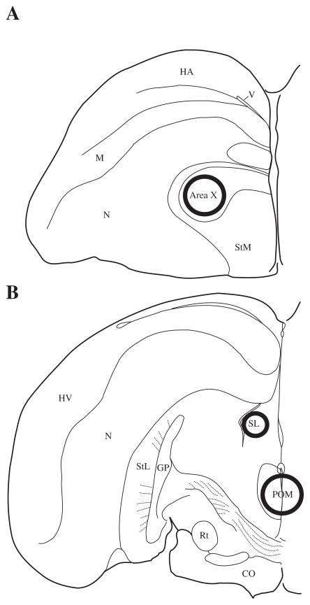
Figure 1.
Location of tissue punches illustrated in one, coronal hemisphere of the starling brain. Approximate punch sizes and locations are represented by circles centered in Area X (1.25mm diameter), POM (1.25mm diameter), and LS (0.75mm diameter). Abbreviations: HA = hyperpallium apicale; M = mesopallium; N = nidopallium; StM = striatum mediale; StL = striatum laterale; GP = globus pallidus; Rt = nucleus rotundus; OC = optic chiasm.

