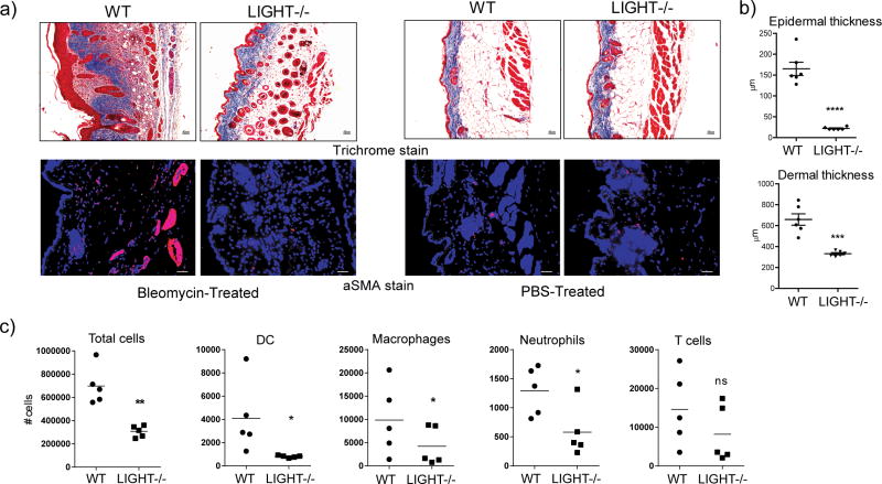
Figure 2. LIGHT-deficient mice exhibit decreased skin fibrosis induced by bleomycin.
WT and LIGHT−/− mice were administered 0.2U bleomycin/mouse. Mice were sacrificed on day 7. (a) Skin fibrosis was assessed by analyzing trichrome (mag. 10x) and aSMA stained sections (mag. 20x). Dashed line delineates the basement membrane. Scale bar = μm. (b) Dermal and epidermal thickness was quantitated. Values from individual tissues from 6 mice. Data are representative of 6 experiments. ***, p < 0.01; ****, p < 0.001. (c) Total CD45+ cells, dendritic cells (DC), neutrophils, macrophages, and T cells, were enumerated in skin samples. Values are from individual tissues from 5 mice.

