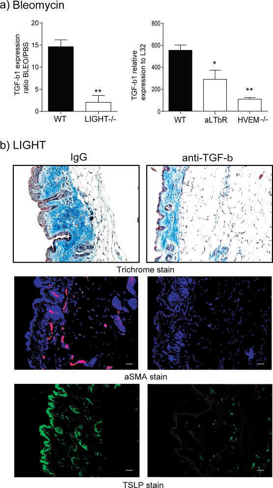
Figure 5. TGF-β is required for LIGHT driven skin fibrosis.
(a) WT, LIGHT−/−, HVEM−/−, and WT mice treated with anti-LTβR, were challenged with intratracheal bleomycin, and after 7 days skin expression of TGF-β1 mRNA was assessed. Values are mean ± SEM of 3 to 4 mice per group. (b) WT mice, treated with control IgG or anti-TGF-β, were administered 10 μg of rmLIGHT on days 1 and 2. Skin fibrosis and TSLP expression was assessed as before on day 3. Dashed line delineates the basement membrane. Scale bar = μm. Data are representative of 3 individual mice analyzed per group.

