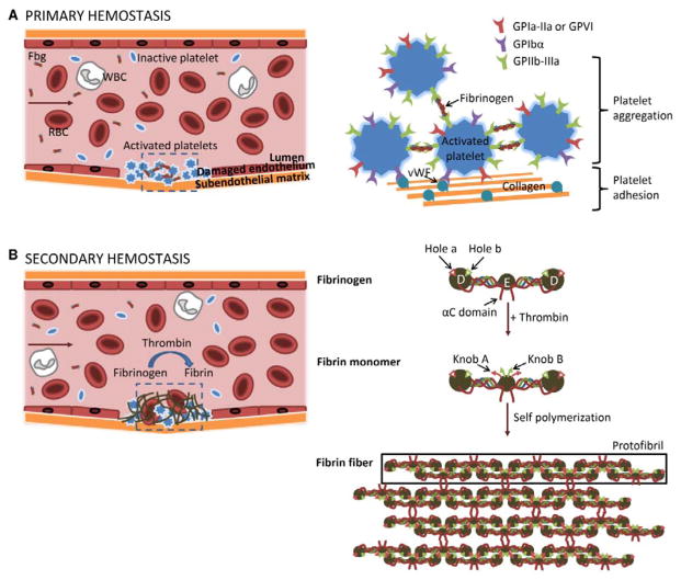
Figure 1.
Schematic of the clotting process showing initial platelet plug formation during primary hemostasis (A) followed by fibrin formation during secondary hemostasis (B). During primary hemostasis, circulating platelets adhere to the subendothelial matrix through binding of the platelet receptors GPIbα and GPIa-IIa/GPVI to von Willebrand Factor (vWF) and collagen, respectively. Upon platelet activation, GPIIb-IIIa goes through a conformational change which enables it to bind fibrinogen. Platelet aggregation is induced by multiple platelets binding the same fibrinogen molecule. During secondary hemostasis, activated thrombin enzyme cleaves the N-termini of Aα (red) and Bβ (green) polypeptide chains, revealing knob A and knob B peptide domains, respectively. Knobs A and B interact with holes a and b in the D nodules of other fibrin monomers to form half-staggered, double-stranded protofibrils. Non-specific interaction between αC domains causes lateral aggregation of protofibrils to form fibrin fibers.

