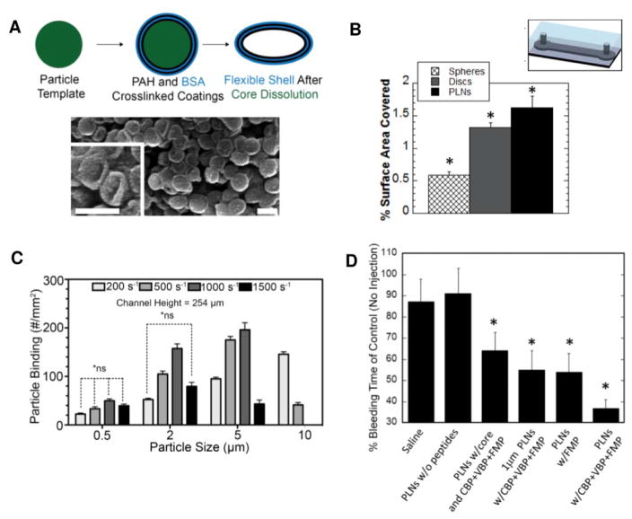
Figure 3.
Data showing effects of particle size, shape, and flexibility on adhesion to surfaces under flow and in vivo hemostatic function. (A) Synthesis of platelet-like nanoparticle (PLN) without targeting peptides. SEM imaging confirmed collapse of the flexible PAH-BSA shell after dissolution of the polystyrene core. Scale bar = 200 nm [reproduced with permission from 45]. (B) Quantification of adhesion to anti-OVA antibody-coated microfluidic chambers of OVA-covered spheres, rigid discs, and PLNS at fixed particles size (200 nm). *Denotes statistical difference (P < 0.05) from all other groups [reproduced with permission from 45]. (C) Quantification of adhesion to endothelialized microfluidic chambers of sLea spherical particles of various sizes under a range of shear rates [reproduced with permission from 46]. (D) Bleeding times after intravenous injection of 15 mg/kg PLN formulations followed by tail transections in Balb/c mice. *Denotes statistical difference (P < 0.05) from saline and PLNs without peptides [reproduced with permission from 45].

