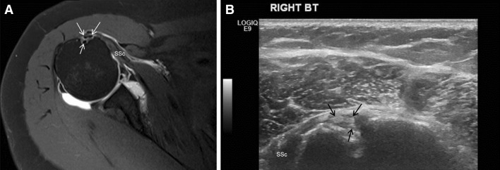
Fig. 1.

a Axial T1-weighted fat suppressed direct MR arthrogram of the right shoulder demonstrates variant long head biceps tendon anatomy consisting of three tendons within the region of the intertubercular sulcus (white arrows). For reference, the subscapularis muscle (SSc) is seen medially attaching to the lesser tubercle. b Short axis US examination of the shoulder demonstrates the same variant long head biceps tendon anatomy (black arrows) in the intertubercular sulcus. The subscapularis muscle (SSc) is again seen medially
