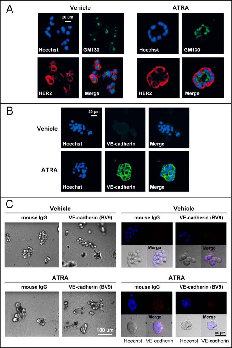
FIGURE 2.
ATRA induces the formation of organized structures reminiscent of the mammary gland epithelium in three-dimensional cultures of SKBR3 cells. SKBR3 cells were grown in a three-dimensional matrix of Matrigel for 5 days and subsequently treated with ATRA (1 μm) or vehicle (ethanol) for 48 h. A, at the end of the treatment, confocal immunofluorescence analysis was performed for the membrane protein HER2 and the Golgi marker GM130. Cell nuclei were stained with Hoechst. The results are representative of three independent experiments. B, SKBR3 cells were grown and treated as indicated above. At the end of the treatment, cells were subjected to immunofluorescence analysis with anti-VE-cadherin antibodies. The cells were counterstained with Hoechst to visualize cell nuclei. The results are representative of three independent experiments. C, SKBR3 cells were grown as detailed above before treatment with ATRA (1 μm) or vehicle (ethanol) for 24 h. Seventeen hours after ATRA administration, cells were incubated with a blocking anti-VE-cadherin monoclonal antibody (BV9) or control IgG, as indicated. Left, bright field images at the indicated low magnification. Right, high magnifications of bright field images are shown in the bottom left panels, whereas the top two confocal images were obtained by immunofluorescence analysis of the same microscopic fields after challenge with anti-VE-cadherin antibodies (top right panels) and staining of the cell nuclei with Hoechst (top left panels). Merged images of the bright field and the confocal fluorescence-labeled fields are illustrated in the bottom right panels (Merge).

