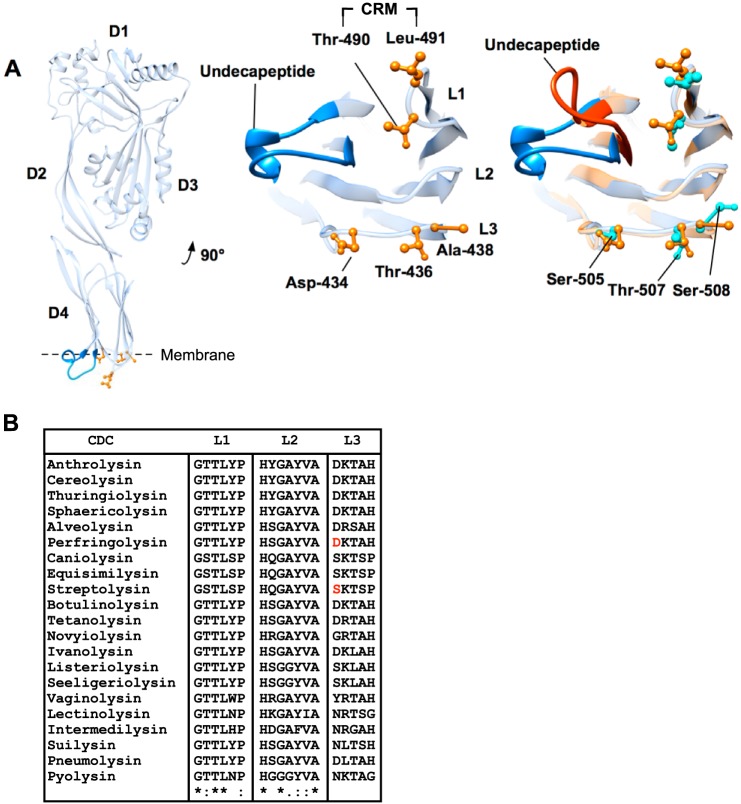
FIGURE 1.
Domain 4 loop structure of PFO and SLO. A, ribbon representation of monomeric PFO (left panel), magnified domain 4 containing the CRM, loops 1–3, and the conserved undecapeptide (center panel). PFO (blue undecapeptide and orange side chains) and SLO (red undecapeptide and aqua side chains) binding structures are overlaid (right panel) to show the analogous SLO residues in L1–L3. Note that the only significant difference in the α-carbon backbone structure of the two toxins is the undecapeptide. Structures were generated using UCSF Chimera (54). B, primary structures of L1–L3 for several known CDCs. The entire primary structures of all the CDCs were aligned using ClustalW (55) to identify the loop regions. Alignment symbols: * = conserved; : = strongly conserved group;. = weakly conserved. The absence of a symbol indicates non-conserved residues. PFO Asp-434 and SLO Ser-505 are highlighted in red in the L3 sequence.

