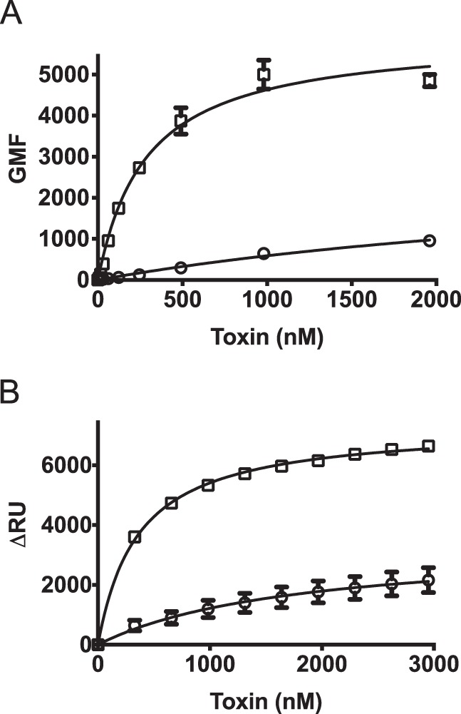
FIGURE 2.

PFO and SLO exhibit different membrane binding parameters. Binding of wild-type SLO (squares) and PFO (circles) was compared on mouse C2C12 myocyte cells by flow cytometry (A) and on cholesterol-rich liposomes by SPR (B). The standard error from at least two separate sets of experiments is shown.
