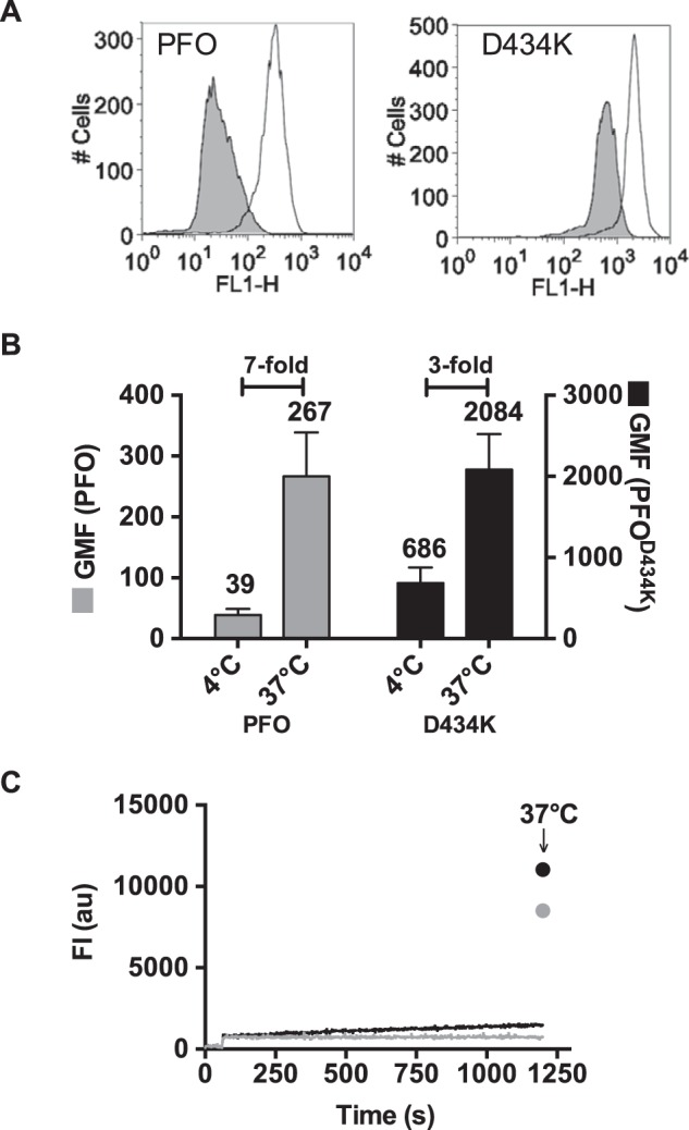
FIGURE 5.

β-Barrel insertion into biological membranes is decreased in PFOD434K. Fluorescence emission of an NBD probe attached to cysteine-substituted Ala-215 in TMH1 of PFO and PFOD434K was measured on C2C12 cells. The efficiency of membrane insertion of the β-barrel can be measured by comparing the NBD fluorescence emission intensities at 4 °C (β-barrel remains uninserted) and 37 °C (β-barrel is inserted) by flow cytometry (5). NBD-PFOA215C and NBD-PFOA215C:D434K were incubated with C2C12 cells for 30 min at 4 °C, and NBD emission of bound toxin was measured by flow cytometry (A, shaded histograms). At 4 °C minimal membrane insertion of the β-barrel occurs, therefore Ala-215 remains in an aqueous environment (see Ref. 24 and C, below). The same samples were then incubated at 37 °C for 15 min to allow for TMH insertion (Ala-215 is inserted into the bilayer), and the fluorescence intensity was measured again (A, open histograms). B, shown are the geometric means of the emission intensities (FLH-1) of bound NBD-PFOA215C and NBD-PFOA215C:D434K. The fold-increases in NBD intensity after the temperature was shifted to 37 °C (due to the positioning of the NBD probe in to the bilayer) is indicated. Mean ± S.E. of two experiments are shown. C, TMH insertion of wild-type PFO (gray) and PFOD434K (black) with an excess of cholesterol-rich liposomes was measured over time at 4 °C for 20 min. The same samples were then incubated at 37 °C for 15 min to allow the β-barrel to insert and the emission of NBD was determined again (arrow).
