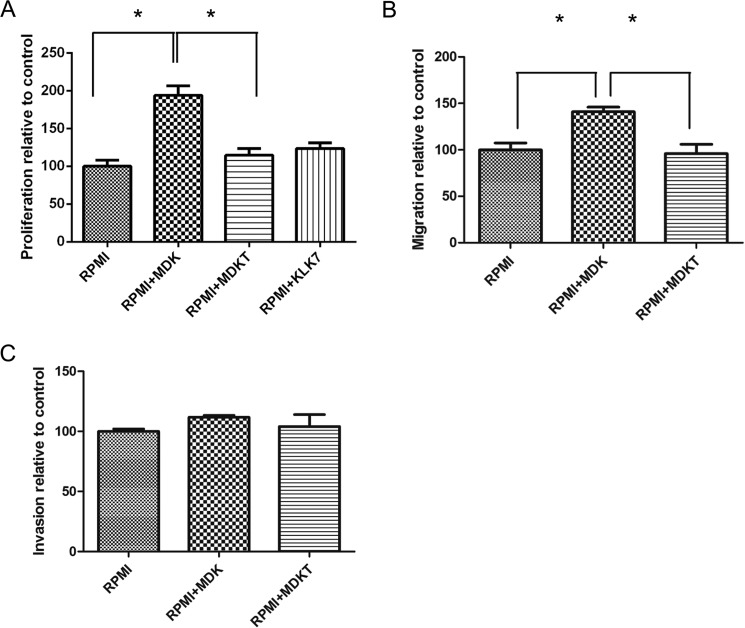
FIGURE 7.
Functional studies of MDK and KLK7-treated MDK (MDKT) in the WM9 cell line. A, proliferation of WM9 cells stimulated with MDK or MDKT. WM9 cells were cultured for 72 h in RPMI1640 medium without additive (RPMI), with 100 ng/ml midkine (RPMI+MDK), or with KLK7-treated midkine (RPMI+MDKT), or 1 ng/ml KLK7 alone. MDKT-truncated form was generated by preincubation of MDK with KLK7 (100:1 ratio) at 37 °C for 3 h. The proliferation rate of cells in RPMI alone was considered as 100%. The data are presented as the means ± SEM (n = 6, p < 0.0001). B, the WM9 cell migration assay. WM9 cells were cultured in RPMI serum-free medium without additive, with 100 ng/ml MDK, or with 100 ng/ml MDKT. The migration rate of cells in RPMI alone was considered as 100%. p = 0.0114. C, WM9 cell invasion assay. WM9 cell were cultured in RPMI serum-free medium without additive, with 100 ng/ml MDK, or with 100 ng/ml MDKT. The invasion rate of cells in RPMI alone was considered as 100%. p = 0.4175. *, p < 0.05. For discussion see “Results.”

