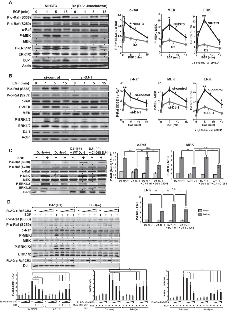
FIGURE 4.
Activation of c-Raf by DJ-1 in EGF-treated cells. A, NIH3T3 and its DJ-1 knockdown D2 cells were cultured in a medium with 0.1% calf serum for 6 h and treated with 50 ng/ml EGF for 1, 5, and 15 min. Proteins extracted from the cells were analyzed by Western blotting with anti-phospho-c-Raf (Ser-338), anti-phospho-c-Raf (Ser-259), anti-c-Raf, anti-phospho-MEK, anti-MEK, anti-phospho-ERK1/2, anti-ERK1/2, anti-DJ-1, and anti-actin antibodies. Intensities of bands were quantified. The number of experiments (n) is 4. Significance: *, p < 0.05; **, p < 0.01. B, HeLa S3 cells were transfected with siRNA-targeting DJ-1 (si-DJ-1) or with nonspecific control RNA (si-control). Forty eight h after transfection, the cells were cultured in a medium with 0.1% calf serum for 6 h and treated with EGF for 1, 5 and 15 min, and expression levels of proteins were analyzed by Western blotting as described in the legend for A. n is 4. Significance: *, p < 0.05; **, p < 0.01. C, DJ-1+/+ cells, DJ-1−/− cells, and DJ-1−/− cells that had been transfected with expression vectors for wild-type DJ-1 or C106S DJ-1 were cultured in a medium with 0.1% calf serum for 6 h and treated with EGF for 3 min, and expression levels of proteins were analyzed by Western blotting as described in A. n is 4. Significance: *, p < 0.05; **, p < 0.01. N.S., nonsignificance. D, DJ-1+/+ and DJ-1−/− cells were transfected with various amounts of expression vectors for FLAG-CR3. Forty eight h after transfection, cells were cultured in a medium with 0.1% calf serum for 6 h and treated with or without EGF for 3 min, and expression levels of proteins were then examined by Western blotting as described in the legend for A. FLAG-CR3 was detected with an anti-FLAG antibody (M2, Sigma). The amounts of expression vectors for FLAG-CR3 transfected to 10-cm dishes were 5, 20, and 40 μg in lanes represented by triangle. n is 4. Significance: *, p < 0.05; **, p < 0.01.

