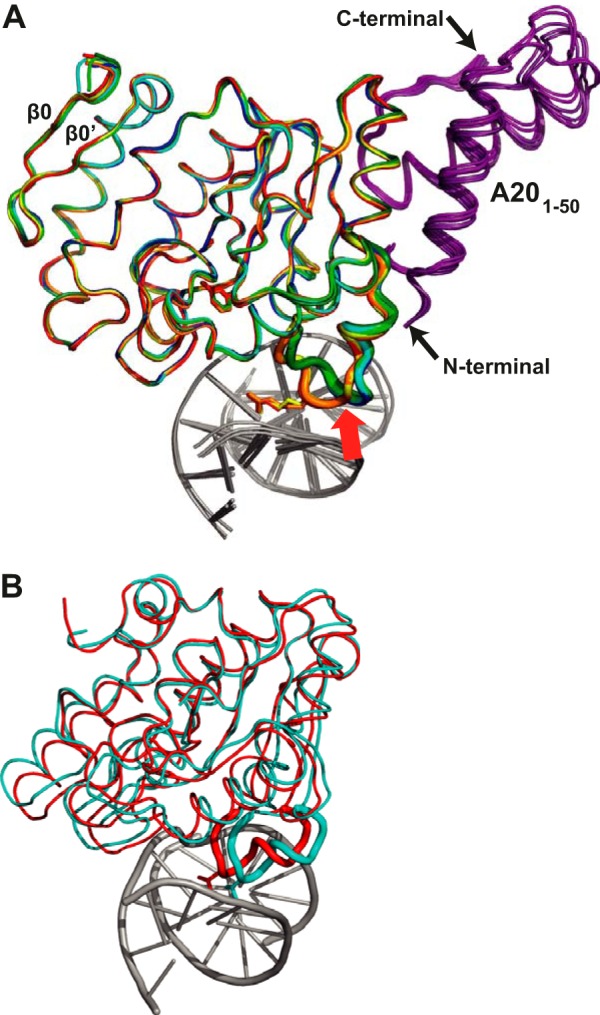
FIGURE 4.

Rigidity of the D4 structure. A, alignment of the backbone of the two distinct D4 apoenzyme structures (dark blue, cyan) (20), of the two D4 structures in complex with uracil (dark green, light green), and of the three D4 structures in complex with DNA (red, orange, and yellow). A201–50 is depicted in violet, and the DNA is in gray. The intercalation loop is highlighted as a thick tube, and the Arg-185 residue is shown in stick representation in the DNA bound structure. This part of the structure is the only part of D4 to show significant rearrangements upon DNA binding (red arrow). The two strands of a D4-specific β-sheet are indicated. B, the corresponding superposition of hUNG in complex with DNA (Protein Data Bank entry 1SSP, red) and the structure of the human apoenzyme (Protein Data Bank entry 1AKZ, cyan).
