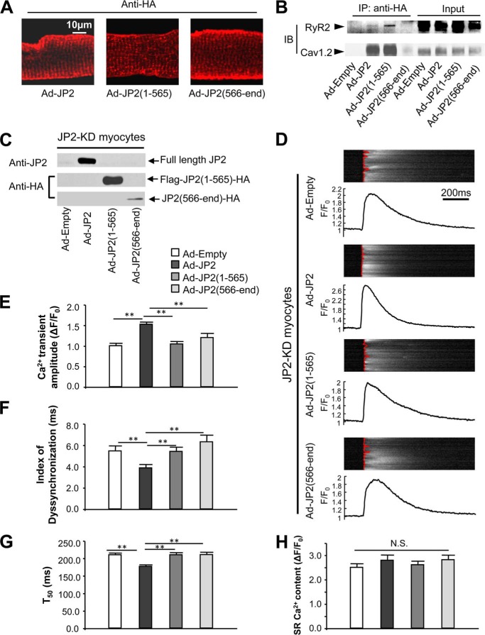
FIGURE 5.
JP2 truncations are nonfunctional in regulating Ca2+ transients. A, an antibody against the HA tag was used to reveal the localization of adenoviral expression of tagged full-length JP2 and truncations, as indicated, in adult wild-type cardiomyocytes. All the three version of JP2 can be localized in the striated pattern. B, full-length JP2 and JP2(1–565) forms complexes with RyR2 and Cav1.2 in vivo. An antibody against HA was used for immunoprecipitation (IP). The RyR2 and Cav1.2 that were pulled down were detected by Western blot analysis. Note that full-length JP2 and JP2(1–565) pulled down both RyR2 and Cav1.2, although JP(566-end) does not form complexes with RyR2 or Cav1.2. IB, immunoblot. C, adenovirus-mediated expression of full-length JP2, FLAG-JP2(1–565)-HA, and JP2(566-end)-HA in JP2-KD) cardiomyocytes. D, representative steady-state Ca2+ transients under 1-Hz field stimulation. The fluorescence intensity (F) of Ca2+ imaging was normalized to the baseline (F0). The red lines overlapping on Ca2+ imaging show the profile of the moment of Ca2+ transient firing on the scanning line in a point-by-point way. A straighter line means better synchronization of the Ca2+ transients. Note that expression of full-length JP2, but not JP2 truncations, improves the amplitude and synchronization of Ca2+ transients. E–G, summary of Ca2+ transient amplitude, index of dyssynchronization (mean absolute deviation of firing time), and duration of 50% decay (T50) (n = 68, 70, 70, and 52 for Ad-Empty, Ad-JP2, Ad-JP2(1–565), and Ad-JP2(566-end), respectively). Only full-length JP2 improves the amplitude, synchronization, and decay of Ca2+ transients. H, summary of SR Ca2+ content, which was assessed by caffeine-induced SR Ca2+ release (n = 17, 17, 17, and 12 for Ad-Empty, Ad-JP2, Ad-JP2(1–565), and Ad-JP2(566-end), respectively). **, p < 0.01 versus indicated groups; N.S., not significant.

