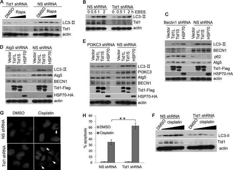
FIGURE 4.
Depletion of Tid1 impairs autophagy induction. A, U2OS cells stably expressing NS shRNA or Tid1shRNA were treated with DMSO or with rapamycin at 0.125, 0.25, 0.5, and 1 μm rapamycin for 6 h. The levels of LC3-II were examined with anti-LC3 blot. B, U2OS cells stably expressing NS shRNA or Tid1shRNA were treated with EBSS at various time points indicated in the figure. The levels of LC3-II were examined with anti-LC3 blot. C–E, HT1080 cells stably expressing NS shRNA, Beclin1 shRNA (C), Atg5 shRNA (D), or PI3KC3 shRNA (E) were transfected with vector, Tid1L-FLAG, Tid1S-FLAG, or HSP70-HA. The levels of LC3-II and other related proteins as indicated in the each figure were examined with specific antibodies. F, U2OS cells stably expressing NS shRNA or Tid1 shRNA were treated with DMSO or 6.25, 12.5, 25, or 50 μm cisplatin for 24 h. The levels of LC3-II, Tid1, and actin were detected with immunoblot using relevant antibodies. U2OS cells stably expressing NS shRNA or mTid1 shRNA were treated with DMSO or 100 μm of cisplatin for 24 h. G, disrupted nucleus was examined with DAPI staining. H, the percentage of cells with disrupted nucleus was statistically analyzed. **, p < 0.01.

