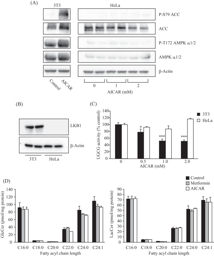
FIGURE 4.
Contribution of LKB1, an upstream kinase of the AMPK pathway, on UGCG activity and GlcCer levels. A, immunoblot analysis showing the protein expression and phosphorylation levels of AMPK α subunit and ACC in 3T3 and HeLa cells. After treatment with AICAR (1 or 2 mm), proteins were extracted from the cells and subjected to immunoblot with antibodies against phospho-Ser-79 (P-S79) ACC, ACC, phospho-Thr-172 (P-T172) AMPK α1/2, AMPK α1/2, and β-actin. B, immunoblot analysis showing a deficiency of LKB1 in HeLa cells. C, intracellular UGCG activity of LKB1-deficient cells. HeLa and 3T3 cells were incubated with several different concentrations of AICAR for 6 h, and then NBD C6-ceramide conjugated with BSA was added to the cells and incubated for 90 min. Fluorescent sphingolipids were extracted from cells and then analyzed by TLC. D, quantification of cellular GlcCer and LacCer levels of LKB1-deficient cells. Lipids were extracted from HeLa cells treated with metformin (8 mm) or AICAR (1 mm) for 16 h and then applied to LC-ESI MS as described under “Experimental Procedures.” Bars, means ± S.D. (error bars) of at least three experiments.*, p < 0.05; ***, p < 0.001.

