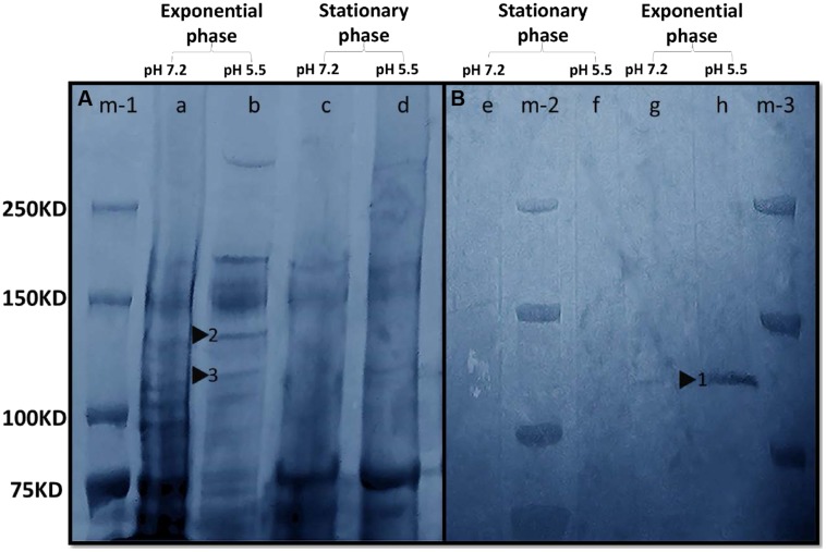
FIGURE 6.
In-gel activity of Mn(II) oxidation enzymes. (A) Gel was stained with Coomassie. Lane m-1, Precision Plus Protein Dual Xtra Standards; Lane a, whole cell protein (pH 7.2, exponential phase); Lane b, whole cell protein (pH 5.5, exponential phase); Lane c, whole cell protein (pH 7.2, stationary phase); Lane d, whole cell protein (pH 5.5, stationary phase). Triangles 2 and 3 indicated the putative Mn(II) oxidizing enzymes with molecular masses of 140 and 120 KDa, respectively. (B) Gel was stained with 200 μM MnSO4. The color of the gel was modified to blue in Adobe Photoshop CC to enhance the clarity of the bands. Lane m-2 and m-3, Precision Plus Protein Dual Xtra Standards; Lane e, whole cell protein (pH 7.2, stationary phase); Lane f, whole cell protein (pH 5.5, stationary phase); Lane g, whole cell protein (pH 7.2, exponential phase); Lane h, whole cell protein (pH 5.5, exponential phase). Triangle 1 indicated the putative Mn(II) oxidizing enzyme with a molecular mass between 120 and 140 KDa.

