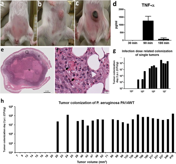
FIG 1.
Infection of transplantable solid tumors in mice. (a) A total of 5 × 10⁵ CT26 cells were injected subcutaneously into the abdomen/flank region. (b) After approximately 10 days, tumors reached a size of between 100 and 150 mm3. (c) Shortly after i.v. P. aeruginosa infection, tumors showed macroscopically visible necrotic areas, like the tumor presented here. (d) Induction of cytokines (exemplified by TNF-α) shortly after bacterial application. TNF-α is most likely the most important cytokine for this model system. (e) Overview of hematoxylin-and-eosin (HE)-stained tumor section colonized by P. aeruginosa. The black box indicates the area that is enlarged in panel f. N indicates the large necrotic area of the infected tumor, and V indicates the remaining viable part of the tumor after bacterial colonization. (A higher-resolution image was added to Fig. S4 in the supplemental material.) (f) Enlargement of the area from the P. aeruginosa-colonized tumor shown in panel e. Tumor-infiltrating neutrophils (black arrows) are identified by their nuclear shape. (g) Efficiency of tumor colonization using different infection doses. Bars represent individual mice. (h) Efficiency of tumor colonization depends on the size of the tumor. Bars represent individual mice.

