
FIG 2.
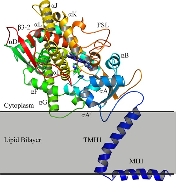
The structure of ScErg11p6×His. The protein is colored from the N terminus to the C terminus with a gradient from blue to red. The heme (purple carbons) and FLC (green carbons) are shown in sticks. For clarity, only some of the α-helices and β-sheets are labeled. FSL, fungus-specific loop.