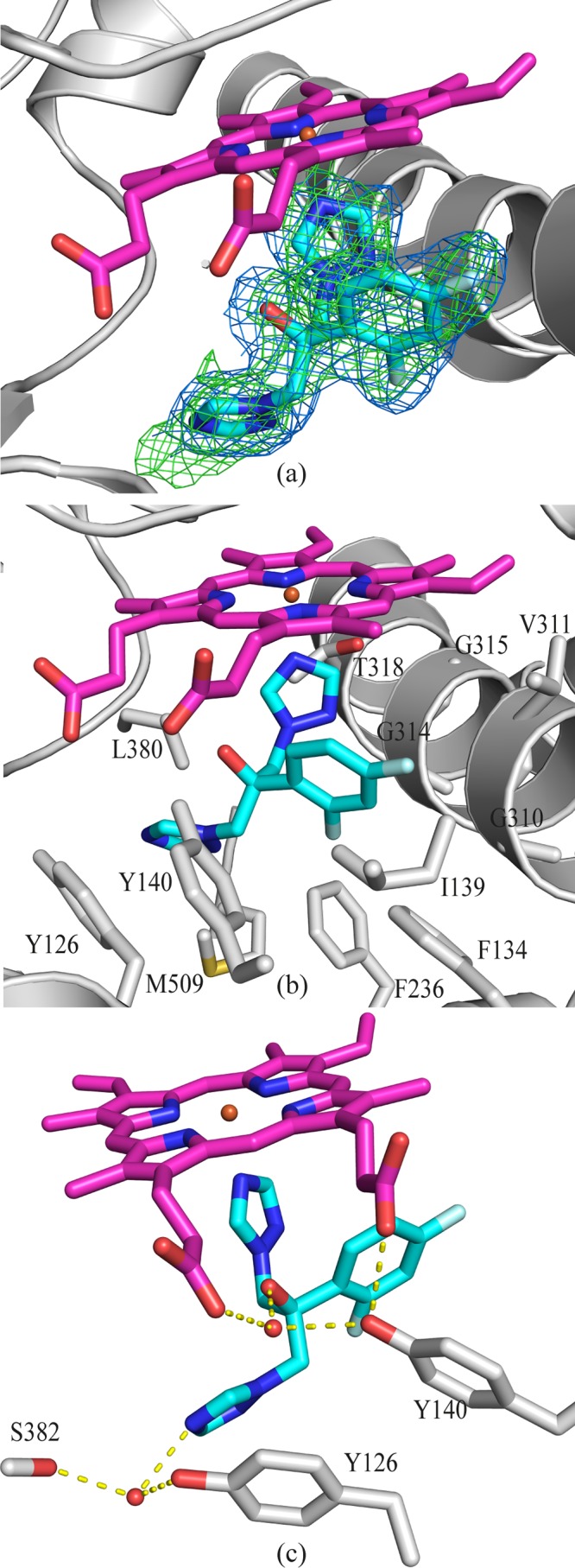
FIG 5.

FLC binding in the active site of ScErg11p. (a) OMIT map for FLC (Fo-Fc map [green mesh] contoured at 3σ; 2Fo-Fc map [blue mesh] contoured at 1σ). The Fo-Fc map was calculated using Fcalc refined from coordinates with no ligand at the active site. The 2Fo-Fc map was calculated following the final refinement. The main chain is indicated in gray, and fluconazole is indicated as sticks, with C atoms in cyan, N atoms blue, O atoms in red, and F atoms in pale blue. The heme is shown as sticks, with C atoms colored magenta. (b) Side chains of amino acid residues within 4 Å of FLC are indicated in gray. The main chain atoms are shown for G310, G314, and G315. (c) Water-mediated hydrogen bonding (yellow dashed lines) between HOH743, FLC, heme, and Y140, as well as between HOH790, FLC, and S382.
