
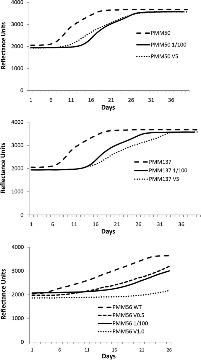
FIG 1.
The lack of PDIM in mycobacteria is associated with glycopeptide susceptibility. Typical fluorometric reflectance results showing mycobacterial cell growth in the absence and presence of 5 (V5), 1 (V1.0), and 0.5 (V0.5) μg/ml vancomycin. (A) Representative growth curves of PMM50 M. bovis BCG (PDIM and PGL deficient) diluted (1/100) or not diluted. The MIC corresponds to a concentration of 5 μg/ml vancomycin. (B) Representative growth curves of PMM137 M. bovis BCG (PDIM deficient, PGL positive) diluted (1/100) or not diluted. The MIC corresponds to a concentration of 5 μg/ml vancomycin. (C) Representative growth curves of PMM56 M. tuberculosis (PDIM deficient) diluted (1/100) or not diluted. The MIC is between 0.5 and 1.0 μg/ml vancomycin.