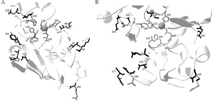
FIG 2.
Ribbon representation of CphA4 and CphA5 superimposed onto CphA structure (Protein Data Bank 1X8G) described by Garau et al. (8). Shown are overviews with all amino acid substitutions labeled for CphA4 (A) and CphA5 (B). In the structure, we have drawn attention to the zinc ion and the residues of Zn1 (N116-H118-H196) and Zn2 (D120-C221-H263) that interact with the zinc ion. The residue in the CphA enzyme is highlighted in gray, whereas the replaced amino acid residues in CphA4 (A) and CphA5 (B) are highlighted in black.

